Medical & Educational Instruments
Head & Brain
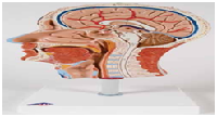
Half Head with Musculature
Representation of the outer, superficial, and internal
(median section) structures of head and neck. Delivered
on removable stand.
22 x 18 x 46 cm; 1.1 kg.
M-C14
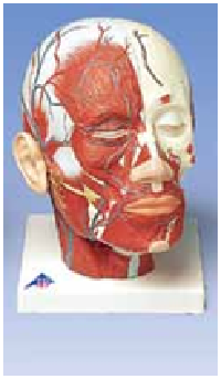
Head Musculature
with Blood Vessels
The same features as M-VB127, plus a
display of the blood vessels.
24 x 18 x 24 cm; 1.2 kg.
M-VB128
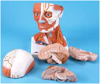
Head and Neck Musculature, 5-part
The model represents the superficial musculature and
deep muscles of the head. The nerves and vessels of the
head are also depicted. The head is dissectible into skull
cap and 3-part brain. Delivered on a removable baseboard.
36 x 18 x 18 cm; 1.8 kg.
M-C05
BESTSELLER
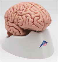
Classic Brain, 5-part
Now with magnetic closures!
This midsagittally sectioned model is an original anatomic cast of a real human brain.
The components of its left half are:
• Frontal and parietal lobe
• Temporal and occipital lobe
• Encephalic trunk
• Cerebellum
13 x 14 x 17.5 cm; 0.49 kg
M-C18
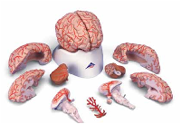
Deluxe Brain
with Arteries, 9-part
This medially divided deluxe brain model shows the brain arteries as welas the detachable basilar artery. On a removable base. Both halves can bdisassembled into:
• Frontal with parietal lobes • Temporal with occipital lobes
• Half of brain stem • Half of cerebellum
14 x 16 x 14 cm; 0.9 kg
M-C20
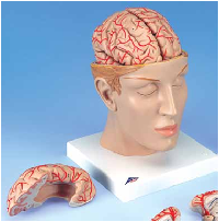
This deluxe brain comes with an
opened head to allow detailed
study of the brain's position in
the skull. The head is horizontally
divided above the skull base.
The brain is divided medially,
with a removable basilar artery.
Both halves can be divided into
frontal parietal lobes, temporal
with occipital lobes, and half of
cerebellum.
15 x 15 x 23 cm; 1.6 kg
M-C25
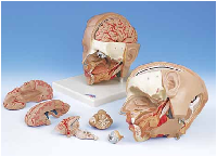
Deluxe Head Model, 6-part
Our most detailed head model! This life-size 6-part head is mounted on a base and
features a removable 4-part brain half with arteries. The eyeball with optic nerve are also
removable. One side exposes the nose, mouth cavity, pharynx, occiput, and skull base.
19 x 23 x 22 cm; 1.0 kg
M-C09/1
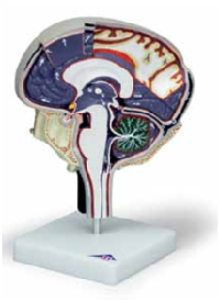
Cerebrospinal Fluid Circulation
Enlarged, detailed model of a section through
the right half of the brain showing the cut pia
mater, arachnoid and dura mater. The model
has the cerebrospinal fluid areas clearly
identified and the direction
of flow indicated
by arrows. Bright
colors to distinguish
important features;
identified in English in an
accompanying product manual.
Mounted on stand.
25 x 18 x 12 cm; 0.9 kg
M-W19027
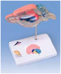
Rat Brain Comparative Anatomy
Enlarged roughly six times, and medially sectioned, the rat brain
model can be disassembled into two halves. The right half of
the color-coded model shows the structures of the cerebrum,
cerebellum, and brain stem. The left half
s largely transparent with a view
of the left lateral ventricle and
hippocampus in the median
section. For comparison, a
natural cast of a rat brain
and a didactic, small-scale
llustration of a human brain
n median section are shown on the base.
Each has the same color coding used for the
various regions.
14 x 10 x 16 cm; 0.24 kg
M-C29
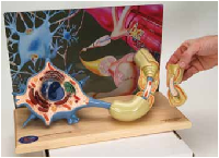
Motor Neuron Diorama
Magnified over 2,500 times, the Diorama reveals details that are normally only seen
through a state-of-the-art electron microscope. This color-coded, three-dimensional
reproduction shows a motor nerve cell situated within a milieu of interacting neurons
and a skeletal muscle fiber. The membranous envelope is cut away from the neuron
to expose the cytological ultrastructure, organelles, and inclusions within the cell
body; branching dendrites, communicating synapses and a myelin-wrapped axon with
node of Ranvier project from the neuronal surface. A section of the axon lifts off to let
you view the tightly wound layers of the enveloping myelin sheath and neurolemma,
as well as the Schwann cell which formed them. Providing perspective and insight
into neuronal function, dendrites of the neuron extend into the background where
they become part of a web-like network of intercommunicating neurons. The axon
converges with the axons from other neurons to form a motor nerve, which ultimately
terminates in a neuromuscular junction or motor endplate. Here, via a cutaway view,
you can observe synaptic vesicles, carrying neurotransmitters, about to stimulate the
muscle fiber to action. Mounted on a wooden base.
M-W42537
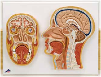
Median and Frontal Section of the Head
The perfect model to learn structure location. Two relief models on baseboard
showing all relevant structures of the human head in great detail.
41 x 31 x 5 cm; 1.1 kg
M-C13