Medical & Educational Instruments
Joints with Ligaments & Muscles
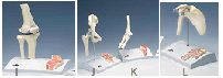
3B Scientific® Mini Joint Series with Cross-Section on Base
Following in the footsteps of their successful larger brothers, these mini-joints have been
reduced to half their natural size but have kept all of their functionality. In addition to the
external anatomical structures, using the superb new joint cross-sections mounted on the
base, educators now have the ability to explain what is happening from “within”.
I. Mini Knee
J. Mini Hip
K. Mini Elbow
L. Mini Shoulder
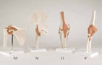
These models provide a graphic demonstration of the anatomy and mechanics
of the major joints, giving your students and patients better understanding. Use
these life-size and fully flexible joints to demonstrate abduction, anteversion,
retroversion, internal/external rotation and more.
M. Flexible Shoulder
16 x 12 x 20 cm
M-A80
Classic Flexible Joint Models N. Flexible Hip
17 x 12 x 33 cm
M-A81
O. Flexible Knee
12 x 12 x 34 cm
M-A82
P. Flexible Elbow
12 x 12 x 39 cm
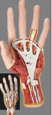
Structural Anatomy of the Hand, 3-part | Internal Structures
Right down to the fingerprints, this full-size model shows
amazing detail. The superficial and internal structures of the
hand including bones, muscles, tendons, ligaments, nerves,
veins, and arteries (superficial and deep palmar arches) are all
present. The palmar aponeurosis and plate of the superficial
flexor tendons are removable. 48 structures are identified in
a multilingual product manual.
Analyze the palmar surface through three increasingly deeper levels:
1st level: palmar aponeurosis 2nd level: exposes the flexor
retinaculum, superficial palmar arch, tendons of the flexor
digitorum, and lumbricales muscles 3rd level: uncovers the
deep palmar arch, and deep layer of muscles, nerves, tendons,
and ligaments.
28.5 x 13 x 6.5 cm; 1.2 kg
M-M18
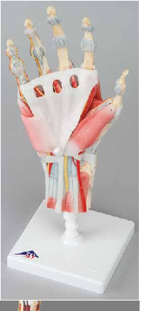
Hand Skeleton with
Ligaments and Muscles
The bones, muscles, tendons,
ligaments, nerves, arteries,
and veins are all featured in
this exquisite 4 part model of the
hand and lower forearm. The dorsal
side shows the extensor muscles as
well as portions of the tendons at the
wrist as they pass under the extensor
retunaculum. The palmar face of the
hand is represented in three layers, the
first two removable to allow detailed
study of the deeper anatomical layer. In
additon clinically important structures
such as the median nerve and superficial
palmar arterial arch can be explored
in detail. The deepest
anatomical layer allows
for study of the intrinsic
muscles and deep
palmar arterial arch in
addition to other details.
33 x 12 x 12 cm; 0.4 kg
M-M33/1
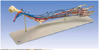
Vascular Arm
Life-size model of the left arm and hand in a semi-flexed position with the
brachial, radial and ulnar arteries and accompanying veins with their radicals
in situ. The complete
blood circulatory system
of the hand is shown on
both palmar and dorsal
surfaces. Comparative sizes of
the various blood vessels are
clearly indicated and facilitate
the study of the blood
circulation in the arm.
Mounted on stand.
66 x 18 x 28 cm, 2.0 kg
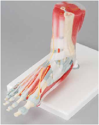
This anatomically detailed model of the foot and lower leg comes with 6 removable
parts for detailed study of the area. The model features not only the bones but also the
muscles, tendons, ligaments, nerves, arteries, and veins. The frontal view features the
extensor muscles of the lower leg. The tendons can be followed as they pass under the
transverse and cruciate crural ligaments all the way to their insertion points. In addition
all tendon sheaths are visible. On the dorsal portion of the model the gastrocnemius
muscle is removable to reveal deeper anatomical elements. The sole of the foot is
represented in three layers; the first layer displaying the flexor digitorum brevis. This
muscle can be removed revealing the quadratus plantae, the tendon of the flexor
digitorum longus, and the flexor hallucis muscle. This
second layer is in turn removable to display even deeper
anatomical details. This model is the best of its kind in
quality and value.
23 x 26 x 19 cm; 1.1 kg
M-M34/1
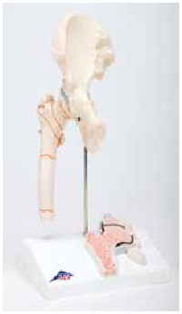
Femoral Fracture and Hip Osteoarthritis
At half natural-size, this model shows the right hip joint of an
elderly person. Also, a frontal section through the femoral
neck is shown in relief on the base. Shown are the femoral
fractures that occur most commonly as well as typical wear
and tear of the hip joint. On stand.
14 x 10 x 22 cm; 0.3 kg
M-A88
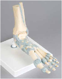
Foot Skeleton with Ligaments
This detailed model displays numerous important
ligaments and tendons including the Achilles
and peroneus longus tendons of the ankle.
The model consists of the foot bone
and lower portions of the tibia and
fibula, including the introsseous
membrane found between
them. All the anatomically
important ligaments and
tendons are shown,
large and small.
23 x 18 x 30 cm; 0.6 kg
M-M34