Medical & Educational Instruments
Skins
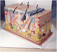
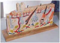
Skin Block Model,
70 times full-size
This distinctive model shows a
section of human skin in three dimensional
form. Individual skin layers are differentiated and
important structures such as hair, sebaceous and sweat
glands, receptors, nerves, erector pili muscles and vessels are
shown in great detail. Mounted on baseboard.
44 x 24 x 23 cm; 3.6 kg
M-J13
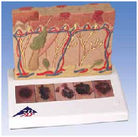
Skin Cancer Model
This 3B Scientific® Pathology model shows 6 different
stages of the malignant melanoma on the front and
back, enlarged 8 times:
• Aggressively healthy malignant cells are found at the surface, within the epidermis
• Malignant cells fill the epidermis, a few invade
the papillary layer
• Malignant cells fill the papillary layer
• Malignant cells invade the reticular layer
• Malignant cells have reached the subcutaneous
fatty tissue, satellite cells approach a vein
In the top view, the individual stages of externally visible
skin changes are shown, allowing for an assessment
according to the “ABCDE” criteria. The sides of the model
show the various levels of invasion into the skin layers
according to Clark (I-V) and the tumor thickness according
to Breslow (in mm). Five original color illustrations on
the base show various types of malignant melanomas.
Mounted on a base.
14 x 10 x 11.5 cm; 0.2 kg
M-J15
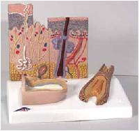
Skin, Hair, and Nail Microscopic Structures
This model shows the microscopic structure of the skin in great detail.
Both hairless and hairy skin structure are shown as well as the different
cell layers of the skin, embedded sweat glands, touch receptors, blood
vessels, nerves, erector pili muscle, and a hair follicle. In
addition to these details, a section of nail is shown on the
base depicting the nail plate, bed, and root. Completing the
skin model is a representation of a hair root with all of its
cellular layers.
10 x 12.5 x 14 cm; 0.35 kg
M-J14
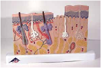
Skin Section, 40 times life-size
The two halves of this relief model show the three layers of
hairy and hairless skin in order to make the differences clear.
In detail with hair follicles, sebaceous glands, sweat glands,
receptor, nerves, erector pili muscles and vessels. Delivered
on base.
24 x 15 x 3.5 cm; 0.2 kg
M-J11
Human Skin Series with Burn Pathologies, 75 times life-size
Six models in one. The front face compares and contrasts the normal
healthy skin from three different body regions; the palm or sole (totally
hairless), the axilla or armpit (sparsely endowed with hair), and the
scalp (completely hirsute). The back of the model illustrates the
progressive severity of injury caused by burns – from the
painful reddening and transitory damage of the first degree
burn, to the blistering, and often permanent damage of the
second degree burn, to the deep charring and permanent
tissue destruction of the third degree burn. 46 features are
coded for identification in an accompanying key. Delivered
on wooden stand.
46 x 25 x 8 cm; 2.75 kg
M-W42533